
Services - MRI
Above, are lumbar spine images of a patient who had undergone back surgery but continued to experience pain. The MRI image on the left was acquired with the patient lying down. It shows a normal alignment of the vertebrae. However, when the patient was scanned in an upright position on the same MRI scanner on the same day (image on the right), a dramatic spinal instability was clearly revealed. This problem, visible only when the patient was scanned upright, would have gone undiagnosed on a conventional, lie-down MRI scanner.
Multi-Position MRI
The Upright MRI is unlike any other scanner because it can scan patients in any position. Because of its true open design, claustrophobic reactions are minimal, larger patients (up to 500lbs) are scanned comfortably and children can be scanned while sitting on a parent's lap. Patients can be scanned sitting or standing while watching a 42" flat screen TV.
The different MRI services offered by DRCTC are:
- Upright MRI
- Open Wide
- Bore High
- Prostate MRI w/CAD
- Breast MRI w/CAD
- MRI Guided Breast Biopsy
- MRI for Thoracic Outlet Syndrome
scheduling
MRI/MRA exams can be scheduled for either the Fort Pierce location or the PORT ST. LUCIE.
Get Scheduling
CAT / CT / CTA Scan
Computed Tomography (CT), also known as a CAT scan, is a sophisticated imaging technique that shows different levels of the anatomy. During CT imaging, the x-ray source rotates around the patient. Each rotation produces a single cross-sectional "slice", like the slices in a loaf of bread. These cross-sectional images are far superior in detail than standard x-rays and greatly enhance a physician's diagnosis.
CT is used to diagnose many conditions. In cancer detection, CT is used to scan for abnormal masses, showing the size and shape of the tumor, its precise location and whether it's solid or hollow. In addition, CT scans can provide valuable information in the detection of abscesses, strokes, head injuries and bleeding inside the skull.
scheduling
Cat Scan exams are performed at our FORT PIERCE & PORT ST. LUCIE location only and take approximately 30 minutes
Get Scheduling
Ultrasound
Ultrasound is a safe, radiation-free imaging procedure that utilizes echoes of sound waves to visualize soft tissue and fluids in the body. It is particularly good in examining the organs in the abdomen and pelvis (liver, gall bladder, pancreas, kidneys, spleen, bladder, uterus, ovaries and testicles). It is also used to evaluate fetal development, certain breast conditions and the thyroid gland.
A modified ultrasound technique, known as Doppler, captures moving blood images of the heart and large blood vessels.
scheduling
Ultrasound exams are performed at our FT. PIERCE & PORT ST. LUCIE location and take approximately 20 minutes.
Get Scheduling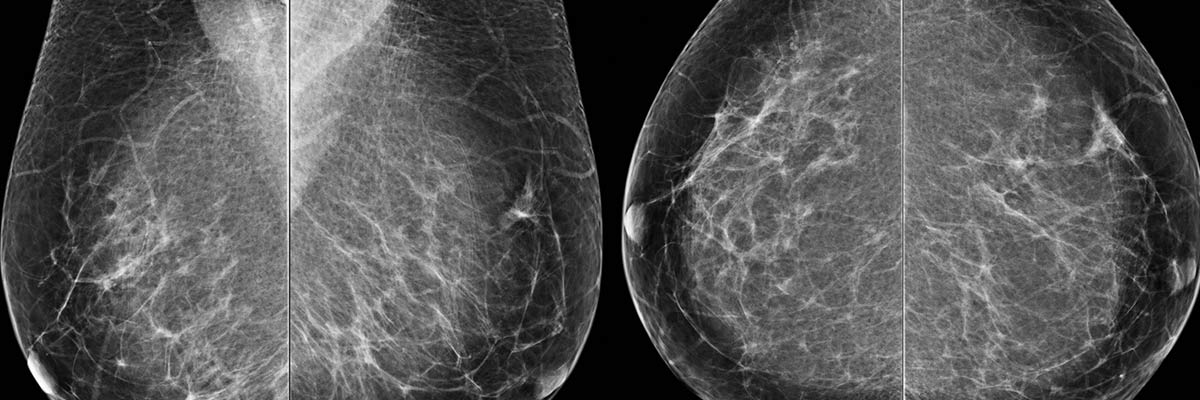
Mammography
At DRCTC, our digital mammography system produces high- quality images that allow for better visualization of breast tissue, aiding in the early detection of breast cancer. Early detection has been shown to dramatically increase the survival rates of women afflicted with this deadly disease.
The Siemens is a state-of-the-art breast cancer detection system designed to improve the quality and comfort of mammograms. This advanced system offers physicians precise viewing options that were never possible with conventional film.
A physician can zoom in, magnify and optimize different parts of the breast tissue, enhancing readability and interpretation of the images. Plus, the images can be sent anywhere in the world to be viewed by another physician.
Compared to conventional film, digital images give better visibility of the breast, particularly near the skin line and chest wall. They�re ideal for women with implants. For women with dense breast tissue, digital is far superior and may involve less radiation than a standard mammography.
In addition, digital exams usually take less than half the time of traditional exams and result in a 20 to 30 percent reduction in callbacks.
scheduling
Mammography exams are also performed at our Fort Pierce location and take approximately 20 minutes.
Get Scheduling
DEXA Scan
DEXA is an advanced technology that safely, accurately and painlessly measures bone density and mineral content in order to determine the risk of developing osteoporosis
Approximately 50% of women over age 50 are at risk of sustaining an osteoporotic fracture. With DEXA, doctors are better equipped to detect and treat bone loss in its earliest stages, so as to prevent the disease or lessen its impact.
At DRCTC, we employ the very latest bone densitometry equipment. During a comprehensive DEXA evaluation, a dual energy beam scans one or more areas, usually the fracture-prone hip or spine. It measures the amount of x-rays that are absorbed by the bones in the body. The two x-ray energies allow the machine to differentiate between bone and soft tissue, giving an accurate estimation of bone density.
Bone Mineral Density (BMD) is calculated and compared to normal BMD values, matched for age and sex, to confirm or exclude osteoporosis. A low BMD may predict the likelihood of developing osteoporosis and can help determine a proactive treatment plan.
scheduling
Dexa Scans are performed at our Fort Pierce location and take approximately 20 minutes.
Get Scheduling
X-ray
Radiography is the use of X-rays to view a non-uniformly composed material (i.e. of varying density and composition) such as the human body. A heterogeneous beam of X-rays is produced by an X-ray generator and is projected toward an object. The density and composition of each area determines how much of the ray is absorbed. The X-rays that pass through are captured behind the object by a digital detector. The detector gives a 2D representation of all the structures superimposed on each other..
scheduling
X-ray exams are performed on a walk-in basis at our Fort Pierce location; except IVP exams which must be scheduled and take approximately 30 minutes.
Get Scheduling
PETCT New
DRCTC is proud to have a mobile PET/CT scanner. This system boasts a wide variety of revolutionary advancements, including being the only Open Gantry Design PET/CT in the marketplace. The system produces the fastest total body imaging time of any PET/CT currently available and the multi-slice CT, allows for highly accurate and precise anatomical mapping.
PET imaging measures the body's metabolic activity. A patient receiving a PET scan is injected with a radioactive isotope which is in the form of glucose. Glucose is metabolized by all living cells in the body. The PET scan images the metabolic activity in the body. In the case of cancer, the cells function in a hyper metabolic state and this is seen on the PET scan.
Therefore PET scanning has become the gold standard in staging and restaging cancers. In cardiovascular and neurological diseases, the PET scan shows areas of increased, diminished or absent metabolic activity, therefore pinpointing abnormalities.
The advantage of PET/CT is that it is a hybrid imaging device, combining the best of seperate technologies. PET is a nuclear medicine imaging device that allows for very sensitive imaging of metabolic and functional processes. CT scanning is very strong in anatomical location. So by combining the units in to one single imaging device, you get the benefits of both technologies. When a patient has a PET/CT scan, the PET will identify areas of altered metabolism which indicate a disease state, and the PET images are fused with the CT images which are taken at the same time, allowing for the precise anatomical location of were the abnormal areas are seen on the PET scan. This gives the physician powerful diagnostic information,and allows the physician to plan the proper and most effective course of treatment.
scheduling
This system boasts a wide variety of revolutionary advancements, including being the only Open Gantry Design PET/CT in the marketplace.
Get Scheduling