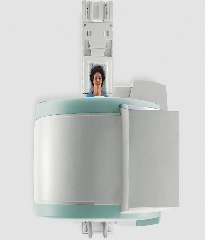
TIM TECHNOLOGY
- Flexibility to meet the needs of a greater patient population
- Accuracy from local to whole body
- Speed – Parallel in all directions for fast exams
- Shortest system length 125 cm combined with 70 cm Open Bore for most head-out exams
- Proven by more than 1.100 installations
- Lightweight coils
- Maximized patient access and dedicated positioning possibilities
- Comprehensive 1.5T application portfolio from routine to advanced applications
(772) 468-7020
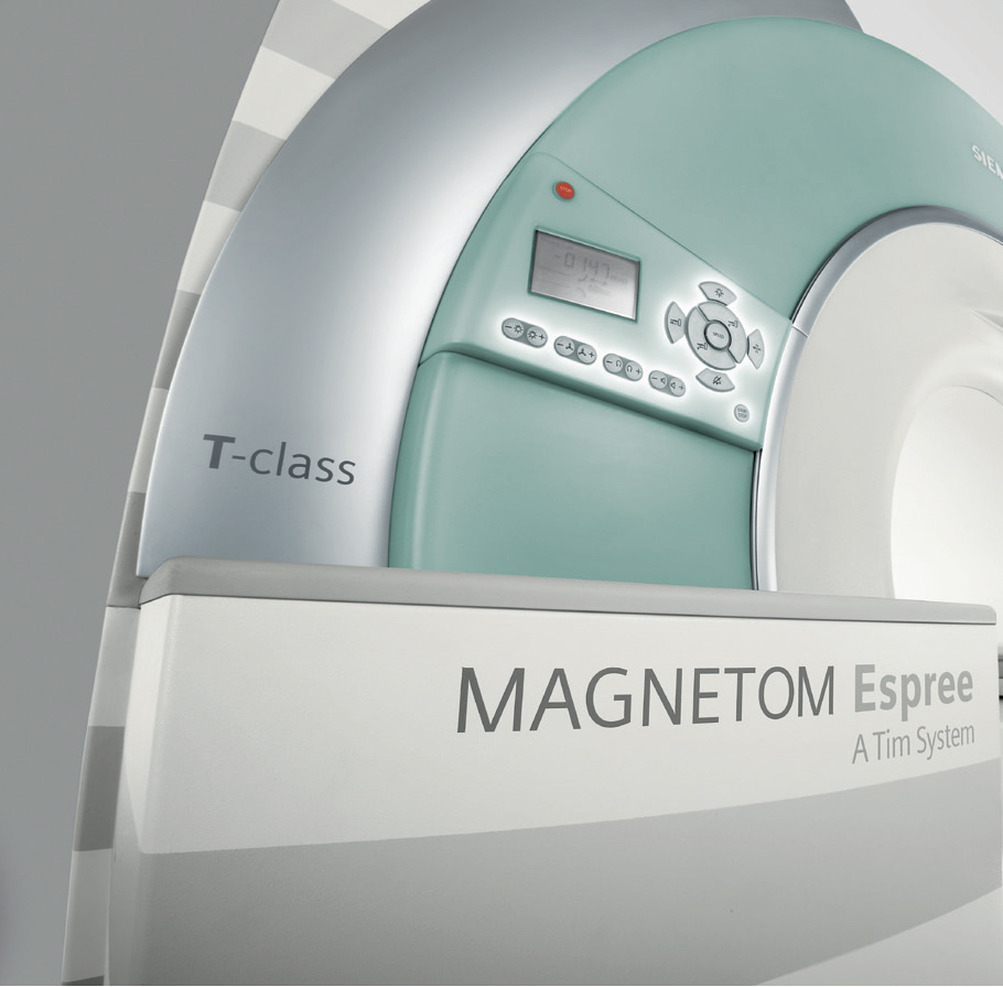
Open High Field 1.5T MRI
2011 S. 25th Street, Suite 106
Fort Pierce, FL 34947
and
Upright MRI
1501 SE Lennard Rd
Port St. Lucie, FL 34952

2 Locations to Serve you!
Open High Field 1.5T MRI
2011 S. 25th Street, Suite 106
Fort Pierce, FL 34947
and
Upright MRI
1501 SE Lennard Rd
Port St. Lucie, FL 34952
(772) 468-7020

Applications


Low Dose Radiation CT Maximum Care Minimum Radiation

Medical Imaging technologies have become essential tools in medicalcare. Clinical images help doctors to diagnose and treat a wide variety of conditions more accurately than ever before. Benefits of imaging include early diagnosis of disease and avoiding unnecessary exploratory surgery. Advances in modern medical imaging technology have made it possible to produce highly accurate images using very low radiation dose.
- Radiation dose reduction is particularly important when imaging children, women of childbearing age, and patients who need multiple X-ray or CT examinations.
- Reducing radiation exposure today means reducing the risks of acute and potential long-term damage.
- The health and safety of our patients is our highest priority.
- Weight limit: 420 lbs.
2 Locations to Serve you!
2011 S. 25th Street, Suite 106
Fort Pierce, FL 34947
and
1501 SE Lennard Rd
Port St. Lucie, FL 34952
(772) 468-7020
Low Dose Radiation 16 Sl ice CT Maximum Care Minimum Radiation
CT is a non-invasive, fast and simple imaging procedure delivering crucial diagnostic information. Technical and Clinical Benefits: Simplifies Imaging Patient friendly design virtually eliminates claustrophobia

CT Excellence in:

Orthopedic Imaging
Exams we offer include : Fracture and Joint Evaluation,Pre- & Post-Surgical Assessment,Spine Procedures, Metal Implants

Urological Imaging
Exams we offer include : Urograms, Morphological or Congenital Defects, Kidney Stone, Vascular Evaluation and Cancer

Vascular Imaging
Exams we offer include : Run-offs, Dissection, Stent Planning & Evaluation, Pre-& Post-Carotid Surgery and CT Angiography

Ear, Nose and Throat Imaging
Exams we offer include : Sinusitis, Mastoiditis, Evaluation of Boney Structures, Inner Ears, Nasopharyngeal Cancer

Pulmonary Imaging
Exams we offer include : COPD, Pre- and Post-Surgical Planning, Pulmonary Embolism, Restrictive Lung Diseases, Malignant or Benign Lesions, CT Angiography, Lung Infections and Abscesses, Pleural Cavity Diseases (e.g. Pneumothorax) and Atelectasis

Pediatric Imaging
Pediatric-specific protocol for minimal radiation dose
Exams we offer include : Trauma imaging, abdominal, vascular disease, respiratory disorders and oncology

Gastro-abdominal Imaging
Exams we offer include : Colon-Polyp Evaluation, Pre-& Post-Surgical Assessment, Abdominal Cancer, Appendicitis and Vascular Evaluation

Neurological Imaging
Exams we offer include : Vascular Assessment, Neuro Infections & Abscesses (e.g.Encephalitis), Stroke Evaluation, Pre- and Post-Surgical Planning, Neural Neoplasms and Spinal Cord Disorders
scheduling
Cat Scan exams are performed at both location only and take approximately 30 minutes.
Get Scheduling
MRI GUIDED BREAST BIOPSY
Breast Magnetic Resonance Imaging (MRI) helps detect breast cancer earlier in patients who are at high risk for breast cancer or who have had breast cancer already. This technique captures multiple cross-sectional pictures of the breast and – through the use of a computer – generates highly detailed 2D and 3D images of the breast and its surrounding tissues for a faster, more accurate diagnosis of breast cancer.
With your comfort in mind, the breast MRI uses a specially designed, removable table that is padded and contoured to fit a woman’s body. The coils can adjust for all sizes of breasts up to FF, and the machine can be entered feet-first, which can be very comforting for claustrophobic patients.
Breast Biopsy
If an abnormal area appears on your mammogram, a breast biopsy may be needed to determine if cancer is present. During a breast biopsy, a needle is inserted into the breast to obtain tissue samples from the area of concern. Done onsite at the Diagnostic Radiology Center of the Treasure Coast, Inc., this procedure can be performed while the patient is sitting or lying down. It requires only a local anesthetic and about an hour of the patient's time.
scheduling
This study is done in fort pierce office.
Get Scheduling
BREAST MRI W/CAD
Computer-aided-detection (CAD) is an automated, efficient way to process and interpret studies and guide interventional procedures. CAD helps to standardize breast MRI study analysis and offers customized reporting, designed to generate highly detailed breast MRI study reports that thoroughly and effectively communicate extent of disease.
Computer-aided-detection (CAD) plays a significant role in improving efficiency, standardization and quality in women's imaging programs. CADstream, the first CAD application designed exclusively for MRI, automates image processing functions and corrects for patient movement during the study. The result is more standardized image processing and analysis, higher quality images and more rapid interpretation of the MRI study. The use of CAD ultimately makes breast MRI more accessible to women who would benefit from this valuable study.
scheduling
Mammography exams are also performed at our Fort Pierce location and take approximately 1 hours.
Get Scheduling
What is MRI of the Prostate?
Magnetic resonance imaging (MRI) is a noninvasive medical test that physicians use to diagnose and treat medical conditions.
MRI uses a powerful magnetic field, radio frequency pulses and a computer to produce detailed pictures of organs, soft tissues, bone and virtually all other internal body structures. MRI does not use ionizing radiation (x-rays).
Detailed MR images allow physicians to evaluate various parts of the body and determine the presence of certain diseases. The images can then be examined on a computer monitor, transmitted electronically, printed or copied to a CD.
The prostate gland is part of the male reproductive system. It is located in front of the rectum and below the bladder, where urine is stored, and surrounds the first part of the urethra, the tube that connects the bladder with the tip of the penis and carries urine and other fluids out of the body. The prostate helps make the milky fluid called semen that carries sperm out of the body when a man ejaculates. Ultrasound and MRI are the most commonly used techniques to image the prostate gland. See the Prostate Ultrasound page for more information.
What are some common uses of the procedure?
The primary indication for MRI of the prostate is the evaluation of prostate cancer. The test is commonly used to evaluate the extent of prostate cancer in order to determine if the cancer is confined to the prostate, or if it has spread outside of the prostate gland.
Occasionally, MRI of the prostate is used to evaluate other prostate problems, including:
infection (prostatitis) or prostate abscess.
an enlarged prostate, called benign prostatic hyperplasia (BPH).
congenital abnormalities.
scheduling
PROSTATE MRI Scans are performed at our Fort Pierce location .
Get Scheduling
DIGITAL X-RAY
Radiography is the use of X-rays to view a non-uniformly composed material (i.e. of varying density and composition) such as the human body. A heterogeneous beam of X-rays is produced by an X-ray generator and is projected toward an object. The density and composition of each area determines how much of the ray is absorbed. The X-rays that pass through are captured behind the object by a digital detector. The detector gives a 2D representation of all the structures superimposed on each other..
scheduling
X-ray exams are performed at our both location; except IVP exams which must be scheduled and take approximately 30 minutes.
Get Scheduling
Ultrasound
Ultrasound is a safe, radiation-free imaging procedure that utilizes echoes of sound waves to visualize soft tissue and fluids in the body. It is particularly good in examining the organs in the abdomen and pelvis (liver, gall bladder, pancreas, kidneys, spleen, bladder, uterus, ovaries and testicles). It is also used to evaluate fetal development, certain breast conditions and the thyroid gland.
scheduling
Ultrasound is performed at both locations
Get SchedulingDEXA SCAN
DEXA is an advanced technology that safely, accurately and painlessly measures bone density and mineral content in order to determine the risk of developing osteoporosis
Approximately 50% of women over age 50 are at risk of sustaining an osteoporotic fracture. With DEXA, doctors are better equipped to detect and treat bone loss in its earliest stages, so as to prevent the disease or lessen its impact.
At DRCTC, we employ the very latest bone densitometry equipment. During a comprehensive DEXA evaluation, a dual energy beam scans one or more areas, usually the fracture-prone hip or spine. It measures the amount of x-rays that are absorbed by the bones in the body. The two x-ray energies allow the machine to differentiate between bone and soft tissue, giving an accurate estimation of bone density.
Bone Mineral Density (BMD) is calculated and compared to normal BMD values, matched for age and sex, to confirm or exclude osteoporosis. A low BMD may predict the likelihood of developing osteoporosis and can help determine a proactive treatment plan.
scheduling
Dexa are performed in both location. This system boasts a wide variety of revolutionary advancements, including being the only Open Gantry Design PET/CT in the marketplace.
Get Scheduling
CT VIRTUAL COLONOSCOPY
DRCTC is proud to have a mobile PET/CT scanner. This system boasts a wide variety of revolutionary advancements, including being the only Open Gantry Design PET/CT in the marketplace. The system produces the fastest total body imaging time of any PET/CT currently available and the multi-slice CT, allows for highly accurate and precise anatomical mapping.
scheduling
CT Virtual Colonoscopy are performed in both locations and must be scheduled.
Get Scheduling
3D TOMO MAMMOGRAPHY
DRCTC is proud to have a mobile 3D TOMO MAMMOGRAPHY. This system boasts a wide variety of revolutionary advancements, including being the only Open Gantry Design 3D TOMO MAMMOGRAPHY in the marketplace. The system produces the fastest total body imaging time of any 3D TOMO MAMMOGRAPHY currently available and the multi-slice CT, allows for highly accurate and precise anatomical mapping.
scheduling
3D TOMO MAMMOGRAPHY are performed at our both location. This system boasts a wide variety of revolutionary advancements, including being the only Open Gantry Design 3D TOMO MAMMOGRAPHY in the marketplace.
Get Scheduling
3D BREAST ULTRASOUND
One of the most promising new technologies is automated 3D ultrasound. Instead of flat, 2D images, this powerful ultrasound technology gives physicians a much more realistic view of the whole breast and its physical structures; providing multiple views — side to side, backand front and more.
Ultrasound evaluation of breast lesions is quick, inexpensive, and does not expose you to potentially harmful ionizing radiation.
scheduling
3D Breast Ultrasound is performed in our Port St. Lucie office only and must be scheduled. This exam can take up to 45 minutes.
Get Scheduling